


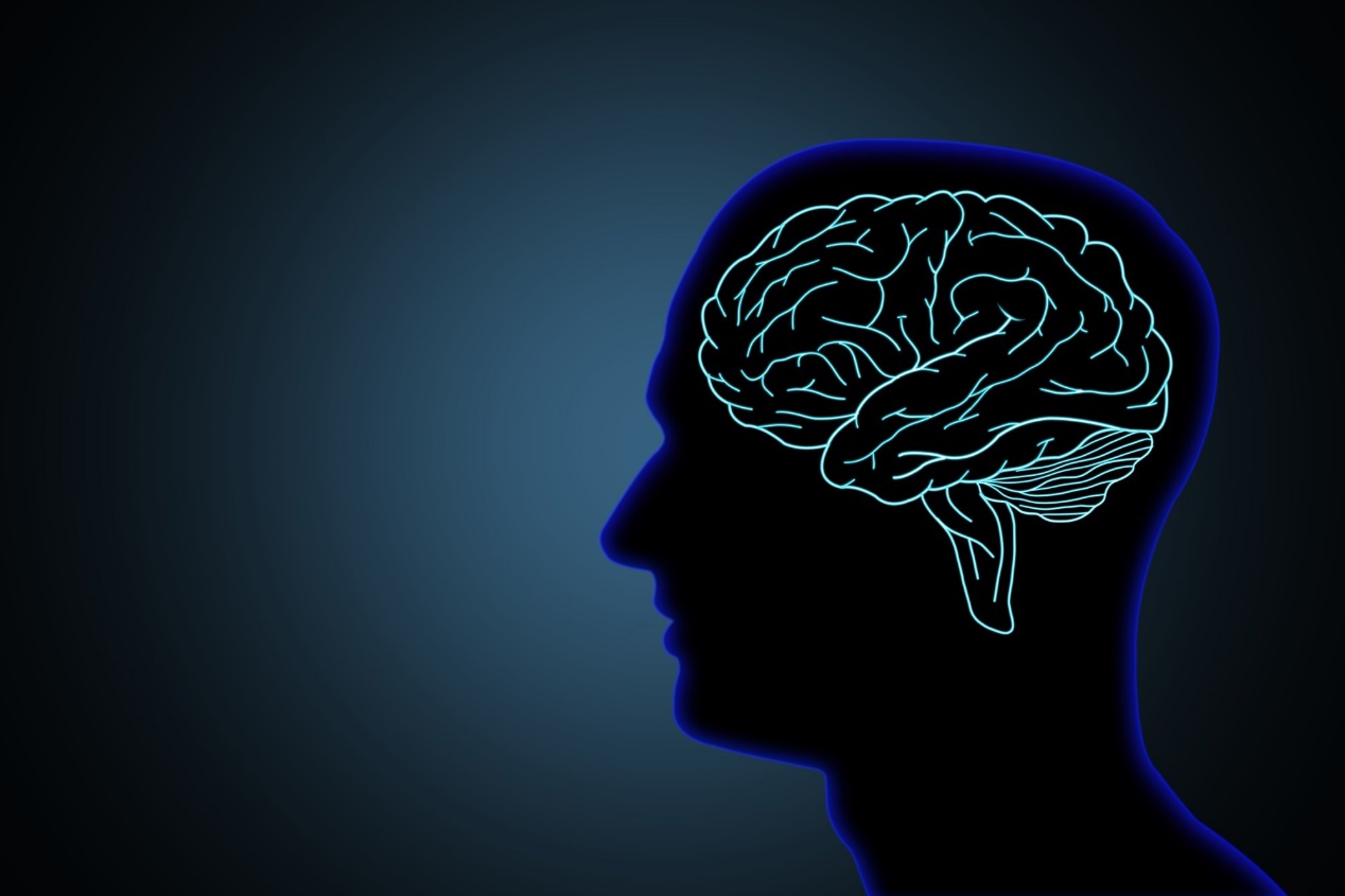
Nature:SARS-CoV-2在人脑组织中的感染、复制和持久性
概要
COVID-19在急性期会引起多器官功能紊乱,少数感染者出现持续症状,这种病状被称为PASC(COVID-19急性后遗症)。然而,后遗症对人体非呼吸系统疾病的负担和SARS-CoV-2从非呼吸系统组织(如大脑)清除的持续时间尚未得到广泛研究。
最近发表在《自然》杂志上的一项研究中,研究人员探索了SARS-CoV-2的细胞趋向性、复制能力、持久性和进化情况,以及受感染组织的相关组织病理学变化。
研究一共分析了44名未接种疫苗和已故COVID-19患者的尸检脑标本,并对11名个体进行了中枢神经系统采样。核酸发现38个样本为SARS-CoV-2阳性,3个样本的SARS-CoV-2血清反应呈阴性,3个样本无法获得血清。此外,研究人员还对病例多处器官进行了采样检测,在大多数病例的脑部区域的所有组织和几种体液(包括血清、玻璃体液和胸膜液)中有检测到了SARS-CoV-2亚基因组核糖核酸。
组织病理学分析结果表明,92%(n=35)的病例死于弥漫性肺泡损伤或急性肺炎,弥漫性肺泡损伤病例呈时间性进展模式,同时观察到心肌浸润,皮质旁和滤泡增生的症状。然而,尽管SARS-CoV-2 RNA在标本体内广泛分布,但在非呼吸道组织中观察到直接SARS-CoV-2细胞病理学或炎症的证据说服力还不够充分。
总体而言,研究结果表明,SARS-CoV-2可以在感染早期感染大脑等非呼吸组织并在其中进行复制,并在症状出现后持续数月(最多230天)。
Study shows SARS-CoV-2 infection, replication and persistence in human brain tissues
In a recent study published in Nature, researchers investigated the cellular tropism, replication competence, persistence and evolution of severe acute respiratory syndrome coronavirus 2 (SARS-CoV-2) in humans, and associated histopathological changes in infected tissues.
Study: SARS-CoV-2 infection and persistence in the human body and brain at autopsy. Image Credit: Surasak_Photo/Shutterstock
Coronavirus disease 2019 (COVID-19) has reportedly caused multiple organ derangements in the acute period, with a few infected individuals developing persistent symptoms, referred to as PASC (post-acute sequelae of COVID-19). However, the non-respiratory disease burden and the duration of SARS-CoV-2 clearance from non-respiratory tissues such as the brain has not been extensively investigated.
About the study
In the present study, researchers mapped and quantified the distribution, proliferation, and cellular tropism of SARS-CoV-2 in non-respiratory human body tissues such as the brain from the acute COVID-19 period to >7.0 months after the onset of symptoms.
Autopsied brain specimens of 44 unvaccinated and deceased COVID-19 patients were analyzed, and extensive CNS (central nervous system) sampling was performed for 11 individuals between April 26, 2020, and March 2, 2021. Polymerase chain reaction (PCR) analysis was performed to confirm SARS-CoV-2 positivity, and 38 samples were found to be SARS-CoV-2-positive. Three samples (P27, P36, and P37) were seronegative for SARS-CoV-2, and sera were not available for three cases (P3, P4, and P15).
Droplet digital PCR (ddPCR) analysis was performed for SARS-CoV-2 N (nucleocapsid) gene quantification, and ISH (in-situ hybridization) analysis was performed to validate the ddPCR results and to determine SARS-CoV-2 cell tropism. The IF (immunofluorescence) and IHC (immunohistochemistry) analyses were performed further to verify the viral presence within human brain tissues.
Further, subgenomic ribonucleic acid (RNA) was detected using real-time quantitative reverse transcription-PCR (RT– qPCR) analysis, and virus isolation experiments were performed using Vero E6 cells to demonstrate proliferation-capable SARS-CoV-2 among tissues of respiratory and another origin. SARS-CoV-2 S (spike) gene variant diversity and distribution were measured using HT-SGS (high-throughput, single-genome amplification and sequencing) analysis for six individuals.
Among the autopsied specimens, 17, 13, and 14 were categorized as early cases, mid-cases, and late cases based on the day of infection (d) at death within 14 days, between 15 days and 30 days, and beyond 31 days, respectively. Further, image analysis on interventricular septal tissues of 16 individuals was performed to assess the association between SARS-CoV-2 N RNA detected by ddPCR analysis and SARS-CoV-2 S RNA detected by ISH analysis. Furthermore, N protein-targeted ISH assays, IF analysis, and IHC-based analyses were performed to validate SARS-CoV-2 detection and distribution in the CNS.
Results
Among the study individuals, 30% were women, with a median age value of 63 years, and 61% of them suffered from at least three comorbid conditions. The median duration between the onset of symptoms to hospital admission and death was six days and 19 days, respectively, and the median post-mortem duration was 22 hours.
SARS-CoV-2 ribonucleic acid was present at 84 anatomical sites in significantly greater amounts among respiratory tissues than other tissues. SARS-CoV-2 RNA levels among early cases, mid-cases, and late-cases were 2.0 log10 nucleocapsid gene copies for every nanogram RNA, 1.4 log10 nucleocapsid gene copies for every nanogram RNA and 0.7 log10 nucleocapsid gene copies for every nanogram RNA, respectively. SARS-CoV-2 ribonucleic acid was detected in the perimortem sera of 11 and one early cases and mid-cases, respectively.
SARS-CoV-2 ribonucleic acid was persistently present in several tissues of late cases, despite being below detectable levels in sera of any case. SARS-CoV-2 ribonucleic acid was identified within the CNS among 91% (n= 10) cases, including across most brain areas evaluated in five (out of six) late cases. SARS-CoV-2 subgenomic ribonucleic acid was detected across all tissues and in several body fluids, including serum, vitreous humor, and pleural fluid.
The subgenomic ribonucleic acid RT-qPCR analysis and ddPCR analysis findings correlated closely for 1,025 specimens, particularly among 369 respiratory samples, 496 early cases, and 302 specimens showing SARS-CoV-2 positivity by RT-qPCR and ddPCR analyses. SARS-CoV-2 was isolated among Vero E6 cells from 45% (n=25) of specimens from the lymph nodes, heart, adrenal gland, gastrointestinal tissues, and ophthalmic tissues of early cases.
In addition, SARS-CoV-2 was isolated from the P38 thalamus among Vero E6-transmembrane serine protease 2 (TMPRSS2)-T2A-angiotensin-converting enzyme 2 (ACE2) cells. HT-SGS analysis of 46 specimens from six individuals did not show any non-synonymous SARS-CoV-2 genomic diversity in respiratory tissues and other tissues for P18, P19, and P27. Among P27 samples, two SARS-CoV-2 haplotypes, each comprising a synonymous mutation, were detected preferentially among non-respiratory tissues such as the mediastinal lymph nodes and the left and right ventricles.
In P38, residue D80F was detected in all 31 respiratory, but none of the 490 cranial sequences and residue G1219V was limited to the cranial variants. Among intraventricular septum tissues, the mean SARS-CoV-2 N gene copies/nanogram RNA correlated significantly with the median SARS-CoV-2 S RNA-positive cells. In the second ISH assay, SARS-CoV-2 RNA and protein were observed in the cerebellum and hypothalamus of P38, basal ganglia of P40, and cervical spinal cord of P42.
The histopathological analysis findings indicated that 92% (n=35) of cases died due to diffuse alveolar injury or acute pneumonia, and the diffuse alveolar injury cases showed a temporal pattern of progression. Myocardial infiltrates, and paracortical and follicular hyperplasia was observed. However, despite widespread SARS-CoV-2 RNA distribution in the body, negligible evidence of direct SARS-CoV-2 cytopathology or inflammation was observed in non-respiratory tissues.
Overall, the study findings showed that SARS-CoV-2 could infect and replicate in non-respiratory tissues such as the brain early in infection and persist for months (up to 230 days) following symptom onset.
Source:
News-Medical
Published on December 16 2022
声明:本站文章版权归原作者及原出处所有。本文章系本站编辑转载,文章内容为原作者个人观点,登载该文章的目的是为了学习交流和研究,并不代表本站赞同其观点和对其真实性负责,本站只提供参考并不构成任何投资及应用建议。
本站是一个学习交流和研究的平台,网站上部分文章为引用或转载,并不用于任何商业目的。我们已经尽可能的对作者和来源进行了告知,但是能力有限或疏忽,造成漏登或其他问题,请及时联系我们,我们将根据著作权人的要求,立即更正或删除有关内容。本站拥有对本声明的最终解释权。

